
Shedding light on the hidden biology of nature, mammal skull identification offers an enduring window into ecology, wildlife tracking, archaeology, and forensic taphonomy. Among all mammalian orders, Rodentia is by far the most diverse and abundant, comprising over 40% of all extant mammal species with more than 2,200 recognized kinds. Because rodents occupy every terrestrial habitat from subterranean tunnels to alpine peaks, encountering their skeletal remains in the field, forest, attic, or within raptor pellets is remarkably common. However, because many species share a generalized ancestral blueprint centered around gnawing and herbivorous or omnivorous feeding, distinguishing one rodent skull from another can challenge even seasoned naturalists and field biologists.
A structured, methodical approach changes everything. Rather than guessing based on ambiguous body dimensions, field-experienced specialists rely on anatomical markers embedded directly within the skull architecture. Whether you are conducting a biodiversity survey, analyzing owl pellets, or resolving an attic mystery, understanding these cranial landmarks allows for rapid, definitive identification. This comprehensive guide explores the foundational anatomy, taxonomic classifications, diagnostic metrics, edge cases, and comparative characteristics necessary to master rodent skull identification.
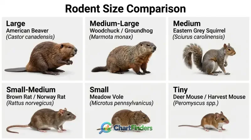
Rodent Skull Identification Chart
- All rodent skulls are defined by a specialized dental layout: a single pair of rootless, continuously growing upper and lower incisors and a prominent, toothless gap known as the diastema.
- Canines and canine-like premolars are entirely absent in all rodent species, separating them instantly from carnivores, omnivores, and insectivores like shrews.
- Skull length serves as the primary sorting metric, breaking the order into large (>150mm), medium-large (90–150mm), medium (45–75mm), small (20–45mm), and tiny (<20mm) categories.
- Snout breadth, the presence or absence of postorbital processes, and molar surface morphology (flat ridges versus rooted cusps or prismatic enamel loops) provide definitive species-level clues.
- Distinguishing rats (Muridae) from squirrels (Sciuridae) requires close examination of zygomatic plate orientation and snout geometry rather than relying solely on body size.
To successfully identify a rodent skull, one must move past general impressions and examine specific diagnostic planes. The following master reference breaks down the primary size classes, cranial features, and dental structures that separate major rodent families encountered in North America and temperate regions.
| Size Class | Typical Skull Length | Representative Species | Key Diagnostic Features | Molar & Dental Morphology |
|---|---|---|---|---|
| Large | > 150 mm (6+ inches) | American Beaver (Castor canadensis) | Massive, heavy bone structure; prominent sagittal crest; robust zygomatic arches; deep orange-pigmented incisors. | Complex ridged flat molars designed for grinding wood and bark; no roots in adults. |
| Medium-Large | 90 – 150 mm | Woodchuck / Groundhog (Marmota monax) | Flattened skull roof; pronounced postorbital processes; robust post-orbital constriction. | Complex rooted cheek teeth with distinct cusps arranged in transverse loops. |
| Medium | 45 – 75 mm | Eastern Grey Squirrel (Sciurus carolinensis) | Shorter, broader snout than rats; arched dorsal profile; distinct postorbital process forming a partial eye socket bar. | Four cheek teeth per quadrant (often with a tiny, vestigial upper premolar that drops out early). |
| Small-Medium | 35 – 50 mm | Brown Rat / Norway Rat (Rattus norvegicus) | Long, narrow snout; lacks postorbital processes; smooth, sloping cranial roof. | Three rows of cusps on molars (tubercles); flat chewing surfaces emerge with wear. |
| Small | 20 – 35 mm | Meadow Vole (Microtus pennsylvanicus) | Blocky, angular skull; specialized prismatic (triangle-shaped) enamel patterns on flat-crowned molars. | Rootless molars growing continuously; complex enamel loops forming triangles. |
| Tiny | < 20 mm (< 0.8 inches) | Deer Mouse / Harvest Mouse (Peromyscus spp.) | Delicate structure; large orbits relative to cranium; distinct notch in the upper incisor profile when viewed laterally. | Biserial cusps in two parallel longitudinal rows; relatively simple chewing surfaces. |
When analyzing a specimen using this matrix, always begin by measuring total condylobasal or greatest length using digital calipers. Once you establish the size tier, you can focus on fine anatomical differences such as molar cusps and zygomatic architecture.
Fundamental Anatomy of the Rodent Skull
Before diving into species-specific traits, you must master the basic cranial architecture shared across the entire order Rodentia. Evolution has shaped the rodent skull into a specialized hydraulic and mechanical lever system optimized for gnawing tough plant material, seeds, wood, and fibrous roots.
The Defining Dental Formula: Incisors and Diastema
The single most reliable feature uniting all rodents is their dentition. Rodents possess a reduced dental formula characterized by the total absence of canines and canine-like premolars. In their place is a wide, empty gap called the diastema, which separates the front gnawing teeth from the rear grinding cheek teeth.
At the front of the mouth sit a single pair of upper and lower incisors. These teeth are remarkable evolutionary innovations: they are open-rooted (ever-growing) and covered in hard, iron-rich orange or yellow-brown enamel on their front surfaces only. Because the softer dentin behind wears down faster than the front enamel during gnawing, the incisors maintain a self-sharpening, chisel-like edge throughout the animal’s life.
Cheek Teeth: Molars and Premolars
Behind the diastema lie the cheek teeth, which include varying numbers of molars and, in some families, premolars. Unlike incisors, cheek teeth are used exclusively for grinding food. Their surface patterns—ranging from simple flat loops of enamel in voles to complex multi-cusped crowns in mice and flat grinding plates in beavers—provide taxonomists with definitive clues for species identification.
Cranial Openings and Muscle Attachments
The power behind a rodent’s bite comes from an extensively modified masseter (jaw-closing) muscle system. Unlike humans or carnivores, where the masseter attaches simply to the lower jaw and cheekbone, rodent masseters extend forward onto the side of the snout. This configuration requires specific cranial modifications:
- Zygomatic Arch: The bony bridge forming the outer edge of the eye socket. Its thickness and the orientation of its front plate (the zygomatic plate) distinguish suborders like Sciuromorpha (squirrel-like), Myomorpha (mouse-like), and Hystricomorpha (porcupine-like).
- Infraorbital Foramen: A critical hole beneath the eye socket through which nerves and blood vessels pass. In many rodents, a portion of the masseter muscle actually originates through this opening, altering its shape from a tiny slit to a massive circular or teardrop-shaped canal.
Classifications and Suborders: Sciuromorpha, Myomorpha, and Hystricomorpha
Mammalogists classify the vast order Rodentia into suborders based primarily on the structure of the masseter muscle and its relationship with the zygomatic arch and infraorbital foramen. Recognizing these structural blueprints helps narrow down any unknown skull:
- Sciuromorpha (Squirrel-like): Characterized by a relatively small infraorbital foramen and a zygomatic plate that is tilted upward. The lateral masseter muscle originates on the zygomatic arch rather than passing through the infraorbital foramen. Representative families include Sciuridae (squirrels, marmots, chipmunks) and Castoridae (beavers).
- Myomorpha (Mouse-like): The largest suborder, comprising mice, rats, voles, hamsters, and jerboas. These skulls feature an enlarged infraorbital foramen shaped like a keyhole or narrow slit, allowing a slip of the medial masseter muscle to pass through and originate on the side of the snout.
- Hystricomorpha (Porcupine-like and Cavy-like): Characterized by a massively enlarged infraorbital foramen through which a large portion of the medial masseter muscle passes. This group includes North American porcupines (Erethizontidae), guinea pigs, and nutria. Their skulls often feature extremely robust zygomatic arches.
Step-by-Step Skull Identification Workflow
When you pick up an unknown skull in the field or laboratory, working through a structured decision tree prevents misidentifications. Follow these sequential steps to narrow down your specimen.
- Verify Order (Is it a Rodent?): Check for the absence of canines and the presence of exactly one pair of large, chisel-like upper incisors and one pair of lower incisors. If there are two pairs of upper incisors (one small pair tucked directly behind the large front pair), the skull belongs to an order called Lagomorpha (rabbits and hares), not Rodentia.
- Measure Total Length: Use digital calipers to measure the greatest length or condylobasal length from the back of the occipital condyles to the front of the nasal bones or incisors. Slot the specimen into one of the primary size tiers (Tiny <20mm, Small 20–35mm, Small-Medium 35–50mm, Medium 45–75mm, Medium-Large 90–150mm, Large >150mm).
- Examine the Snout and Orbits: Look at the top of the skull between the eyes. Do you see bony projections extending backward from the eye sockets (postorbital processes)? If yes, you are likely looking at a squirrel family member (Sciuridae). If the top of the skull is smooth, narrow, and tapers cleanly to the snout without postorbital bars, consider rats, mice, or voles (Muridae/Cricetidae).
- Inspect the Cheek Teeth Morphology: Turn the skull over and examine the chewing surfaces under a hand lens or stereomicroscope. Are the surfaces flat with zig-zag enamel lines (voles/beavers), arranged in distinct rounded tubercles/cusps (mice/rats), or comprised of clean parallel ridges (squirrels)?
- Evaluate Size and Proportion Context: Combine your anatomical findings with geographic range data and local habitat context. For instance, a medium-sized rodent skull found in a suburban attic in North America is almost certainly a Norway rat or roof rat, whereas one found high in an oak canopy is likely a tree squirrel.
Detailed Breakdown by Family and Major Species
To move from broad categories down to individual species, you must examine the subtle anatomical differences that separate major rodent families. Below is an in-depth review of the most commonly encountered rodent groups.
Family Sciuridae (Squirrels, Chipmunks, and Marmots)
Squirrel skulls are instantly recognizable among medium-to-large rodents due to several conservative structural traits:
- Postorbital Process: Sciurids possess a distinct, sharp projection of bone pointing backward from the upper margin of the orbit. In chipmunks and tree squirrels, this forms a clear shelf; in larger marmots, it is robust and flared.
- Snout Proportions: Compared to rats of similar body weight, squirrel snouts are noticeably shorter, wider, and more blunt.
- Zygomatic Plate: The anterior root of the zygomatic arch is tilted upward and lacks the deep vertical notch seen in mouse-like rodents.
Family Muridae and Cricetidae (Rats, Mice, Voles, and Hamsters)
Representing the most speciose group of rodents, these families include familiar pests and vital ecological foundation species:
- Norway Rat (Rattus norvegicus): Skulls range from 40 to 45 mm in length. They feature prominent temporal ridges that run parallel along the braincase (unlike mice, which have smoother, more rounded braincases). The upper incisors curve backward slightly.
- House Mouse (Mus musculus): Much smaller (skull length ~20 mm). The upper incisors possess a distinct, deep notch on their cutting edge when viewed from the side—a critical diagnostic feature separating mice from small shrews (though shrews have pointed insectivore teeth, not chisel incisors).
- Voles (Microtinae): Characterized by blocky, angular skulls and specialized prismatic cheek teeth. Instead of rounded tubercles, vole molars are composed of interlocking triangles of enamel, specialized for grinding abrasive grasses.
Family Castoridae (Beavers)
The American beaver produces one of the most unmistakable skulls in the mammalian world:
- Size and Mass: Skulls routinely exceed 130 to 150 mm in length. The bones are exceptionally thick, dense, and heavily ridged to anchor massive chewing muscles.
- Incisors: Broad, deep, and vividly stained bright orange-red by iron compounds that strengthen the enamel against mineral wear.
- Cheek Teeth: Four large, flat molars per jaw quadrant featuring complex folded enamel patterns that wear down into flat grinding surfaces.
Common Identification Pitfalls and Look-Alikes
Even experienced field biologists occasionally stumble when identifying damaged or juvenile skulls. Awareness of common traps prevents erroneous conclusions.
Rodents vs. Lagomorphs (Rabbits and Hares)
The most frequent error is confusing a small mammal skull with a rabbit. While both have large gnawing incisors and a diastema, lagomorphs possess a second pair of tiny, peg-like incisors hidden immediately behind the primary upper incisors. Rodents possess strictly one pair above and one pair below. Additionally, lagomorph skulls feature a network of highly porous, latticework bone (fenestration) along the sides of the snout that is entirely absent in rodents.
Rodents vs. Insectivores (Shrews and Moles)
Tiny skulls found in owl pellets are sometimes misidentified as mice when they actually belong to shrews. Shrews are insectivores, not rodents. Their skulls lack the single pair of large chisel incisors, featuring instead a complex array of pointed, pigmented teeth designed for grabbing slippery insect prey. , shrews lack a diastema entirely; their teeth form a continuous row.
Juvenile Variation and Subadult Skulls
Young rodents possess proportionately larger eye orbits, thinner cranial bones, and shorter snouts than fully mature adults of the same species. Relying solely on overall skull length without checking dental eruption stages (such as whether the final molar has fully emerged or if sutures are completely fused) can lead to misidentifying a subadult large rodent as an adult of a smaller species.
Frequently Asked Questions
Examine the top of the skull between the eyes. Squirrels possess prominent, backward-pointing bony projections called postorbital processes, and their snouts are relatively short and broad. Rat skulls are smoother across the top, lack postorbital processes entirely, and feature longer, narrower snouts with delicate nasal bones.
No. All rodent skulls completely lack canines and canine-like premolars. This creates a wide, toothless gap known as the diastema between their front gnawing incisors and their rear grinding cheek teeth.
The bright orange or yellow-brown pigment on rodent incisors comes from iron-rich mineral deposits embedded within the hard front enamel layer. This iron reinforcement makes the enamel significantly harder than the softer dentin behind it, ensuring the tooth wears down at an angle that maintains a sharp chisel edge.
While both are small rodents, their cheek teeth differ dramatically. Mouse skulls feature molars with rounded cusps (tubercles) arranged in rows. Vole skulls feature blocky, flat-crowned molars marked by distinctive, geometric enamel loops shaped like interlocking triangles.
Look closely behind the large upper incisors. If you see a second pair of tiny, peg-like “peg teeth” tucked directly behind the front ones, the skull belongs to a lagomorph (rabbit or hare). Rodents have only one pair of upper incisors and completely lack peg teeth.
Conclusion
Mastering rodent skull identification transforms a pile of weathered bone fragments into a rich data source regarding local ecology, predator diets, and biodiversity. By systematically assessing overall skull dimensions, verifying the classic rodent dental formula of single incisors and a diastema, evaluating suborder-specific zygomatic traits, and analyzing fine details like postorbital processes and molar enamel patterns, you can confidently separate everything from tiny meadow voles to massive river beavers. Keep a digital caliper and a good hand lens handy, consult your identification charts, and let the cranial architecture reveal the identity of your specimen.