
The human mouth is a sophisticated biomechanical engine comprising teeth of remarkably diverse shapes, roots, and functions. From the sharp, shearing incisors at the front to the wide, multi-cusped molars at the back, every tooth plays a specialized, non-interchangeable role in mastication, phonetics, deglutition, and facial support. However, translating this biological reality into an unambiguous, universally understood format requires a rigorous standardized framework. Without a reliable mapping tool, clinical notes become ambiguous, treatment errors multiply, and legal or forensic audits fail.
Modern dental practice relies heavily on standardized dental charting to bridge the gap between physical anatomy and digital health records. In contemporary clinical environments—particularly with the widespread adoption of digital odontograms and electronic health records (EHRs)—tooth charts have evolved from simple paper sketches into sophisticated, integrated data hubs. Understanding how these charts are structured, how individual teeth are identified, how different numbering systems operate, and how to avoid critical charting pitfalls is essential for general practitioners, dental specialists, forensic investigators, and healthcare administrators alike.
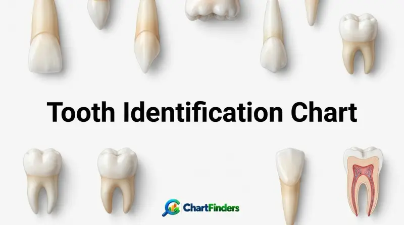
Tooth Identification Chart
- Standardized dental numbering systems (Universal, FDI, and Palmer) eliminate regional and linguistic ambiguity when identifying individual teeth across global clinical, legal, and forensic settings.
- The adult permanent dentition consists of 32 teeth divided into four symmetrical quadrants, while the primary (deciduous) dentition contains 20 teeth devoid of premolars.
- Digital odontograms and electronic dental charts have largely replaced paper records, addressing historical error benchmarks (such as 44% paper audit inaccuracies) to support stringent medico-legal compliance.
- Each tooth class—incisors, canines, premolars, and molars—possesses distinct morphological features, crown contours, cusp numbers, and root configurations that facilitate anatomical identification.
- Systematic charting requires a strict orientation perspective, typically viewing the chart as if looking directly into the patient’s oral cavity (patient’s anatomical right is positioned on the chart’s left side).
At its core, a tooth identification chart functions as a standardized grid or diagram of the human dentition. It provides a visual and alphanumeric shorthand that allows any dental practitioner, radiologist, or forensic odontologist to pinpoint a specific tooth instantly, along with its associated surfaces, restorations, periodontal health status, and pathological conditions. To master tooth identification, one must examine the anatomical classifications of human teeth, understand the mechanics of global numbering systems, evaluate digital odontogram integration, and navigate clinical edge cases.
Anatomical Foundations of the Human Dentition
Human dentition is diphyodont, meaning humans develop two successive sets of teeth throughout their lifetime: the primary (deciduous, milk, or baby) teeth and the permanent (adult) teeth. The transition from primary to permanent dentition is a carefully orchestrated biological process spanning from infancy through early adolescence, characterized by mixed dentition stages.
The primary dentition consists of 20 teeth: 10 in the maxillary (upper) arch and 10 in the mandibular (lower) arch. Each quadrant contains five teeth: a central incisor, a lateral incisor, a canine, a first molar, and a second molar. Notably, primary dentition lacks premolars; these alveolar spaces remain unoccupied until the permanent premolars erupt to replace or supplement the primary molars.
The permanent dentition expands this complement to 32 teeth: 16 in the maxillary arch and 16 in the mandibular arch. Each permanent quadrant contains eight teeth:
- Central Incisor: Broad, flat-edged chisel designed for biting and shearing food. Maxillary central incisors are notably wider mesiodistally than mandibular central incisors.
- Lateral Incisor: Smaller than the central incisor in the upper arch but shares a similar incisal edge function, often exhibiting anatomical variation such as peg-shaped anomalies.
- Canine (Cuspid): Long, single-rooted piercing tooth with a prominent pointed cusp designed for tearing and grasping; possesses the longest root in the human mouth, anchoring the corner of the arch.
- First and Second Premolars (Bicuspids): Transitional teeth possessing two or more cusps (buccal and lingual), combining the shearing action of canines with the grinding action of molars (entirely absent in primary dentition).
- First, Second, and Third Molars: Large, multi-cusped posterior powerhouses designed for crushing and grinding food. The third molars are commonly known as wisdom teeth and exhibit high variability in eruption and morphology.
| Tooth Class | Primary Quantity | Permanent Quantity | Primary Function | Key Anatomical Features |
|---|---|---|---|---|
| Incisors | 8 (4 Maxillary, 4 Mandibular) | 8 (4 Maxillary, 4 Mandibular) | Biting, cutting, shearing | Chisel-shaped crowns, single roots, straight incisal edges |
| Canines | 4 (2 Maxillary, 2 Mandibular) | 4 (2 Maxillary, 2 Mandibular) | Tearing, piercing, gripping | Single prominent pointed cusp, longest roots in the mouth |
| Premolars | 0 | 8 (4 Maxillary, 4 Mandibular) | Grinding, tearing transition | Two or more cusps, prominent buccal and lingual cusps, single or bifurcated roots |
| Molars | 8 (4 Maxillary, 4 Mandibular) | 12 (6 Maxillary, 6 Mandibular) | Crushing, grinding | Large rectangular crowns, multiple cusps (4-5), multiple roots (2-3) |
The table above outlines the quantitative and functional breakdown of human teeth. As demonstrated, the fundamental distinction between primary and permanent arches lies in the inclusion of premolars and the addition of third molars in the permanent set.
Global Tooth Numbering Systems Explained
Because verbally describing a tooth (e.g., “the upper right second molar”) can be cumbersome and prone to misinterpretation across different languages and clinical specialties, international dentistry has established three primary standardized notation systems. A comprehensive tooth identification chart must account for or translate between these standards.
1. The Universal Numbering System
Widely utilized in the United States and favored by many general dental practices, the Universal Numbering System assigns a unique number from 1 to 32 for permanent teeth and capital letters (A through T) for primary teeth.
- Permanent Teeth: Numbering begins at the upper right third molar (Tooth #1), moves across the maxillary arch to the upper left third molar (Tooth #16), drops down to the lower left third molar (Tooth #17), and moves across the mandibular arch to the lower right third molar (Tooth #32). This follows a continuous loop from the operator’s perspective facing the patient.
- Primary Teeth: Utilizing capital letters, the upper right second molar is designated as ‘A’, moving across to the upper left second molar as ‘J’, dropping to the lower left second molar as ‘K’, and ending at the lower right second molar as ‘T’.
2. The FDI World Dental Federation Notation (ISO 3950)
Endorsed by the World Health Organization and the International Association for Dental Research, the FDI system uses a two-digit code. This system is globally prevalent in specialist journals, international clinical trials, and European dental practices.
- Quadrant Designation (First Digit): The mouth is divided into four quadrants. For permanent teeth, 1 represents the upper right, 2 is the upper left, 3 is the lower left, and 4 is the lower right. For primary teeth, the numbers are 5 (upper right), 6 (upper left), 7 (lower left), and 8 (lower right).
- Tooth Designation (Second Digit): Numbered 1 through 8 for permanent teeth (starting from the central incisor outward to the third molar) and 1 through 5 for primary teeth.
- Example: Tooth 11 translates to the Permanent Maxillary Right Central Incisor. Tooth 36 represents the Permanent Mandibular Left First Molar.
3. The Palmer Notation Method
Commonly used in orthodontics and pedodontics, the Palmer system uses a symbol (a grid bracket) to indicate the quadrant, combined with a number or letter representing the tooth.
- A vertical line and horizontal line intersect to represent the midline and the occlusal plane, framing the tooth number.
- Permanent teeth are numbered 1 through 8 starting from the central incisor to the third molar in each quadrant.
- Primary teeth are designated with letters A through E or roman numerals I through V within the quadrant brackets.
| System Name | Primary Dentition Format | Permanent Dentition Format | Primary Application Region |
|---|---|---|---|
| Universal Numbering System | A through T (Upper Right #A to Lower Right #T) | 1 through 32 (Upper Right #1 to Lower Right #32) | United States, general dentistry |
| FDI (ISO 3950) Notation | Two-digit codes: Quadrants 5–8 (e.g., 51 to 85) | Two-digit codes: Quadrants 1–4 (e.g., 11 to 48) | Global, Europe, research, specialist journals |
| Palmer Notation Method | A through E or I through V within quadrant brackets | 1 through 8 within quadrant brackets | Orthodontics, pedodontics |
The Evolution of Digital Odontograms in Modern Practice
Historically, dental charts were hand-drawn sketches on paper folders. While functional, paper-based charts suffered from a notorious vulnerability: legibility issues, missing updates, physical degradation, and a significant audit failure rate. Contemporary clinical and forensic research highlights that paper records struggle to withstand rigorous legal and insurance audits, with historical error benchmarks frequently exceeding 40% in unstructured paper environments.
The introduction of digital odontograms has revolutionized modern clinics by providing interactive, standardized, and tamper-evident graphic interfaces. A digital tooth identification chart offers several distinct advantages:
- Standardized Data Entry: Clinicians click directly on specific tooth surfaces (mesial, distal, buccal, lingual, occlusal/incisal) to record restorations, caries, crowns, missing teeth, and periodontal pockets without handwriting discrepancies.
- Cross-Platform Compatibility: Digital charts integrate with practice management software, allowing instant sharing between general dentists, endodontists, orthodontists, and maxillofacial surgeons without transcription errors.
- Legal and Forensic Defensibility: Every entry is timestamped and tracked via immutable audit logs, creating a secure legal record that satisfies regulatory standards and insurance compliance reviews.
- Visual Analytics: Advanced dental software can analyze historical chart data to highlight disease trends, treatment acceptance rates, and longitudinal changes in oral health.
Step-by-Step Guide to Reading and Navigating a Tooth Chart
Accurately interpreting a tooth identification chart requires a methodical approach. Whether examining a physical chart or navigating a digital odontogram, the following step-by-step framework ensures zero misidentification:
- Establish Anatomical Orientation: Confirm whether the chart is displayed from the “operator’s view” or the “patient’s view.” Standard dental charts display the patient’s right side on the left side of the page or screen (mirroring looking into a mirror or facing the patient).
- Identify the Dentition Type: Check whether the chart references primary (20 teeth, letters A–T or FDI 51–85) or permanent (32 teeth, numbers 1–32 or FDI 11–48) dentition.
- Locate the Midline: Find the vertical line separating the left and right halves of the dental arches. All numbering or lettering systems originate from this central anatomical boundary.
- Determine the Arch: Differentiate between the maxillary arch (upper jaw) and the mandibular arch (lower jaw).
- Trace the Tooth Coordinates: Apply the specific numbering system in use (Universal, FDI, or Palmer) to trace outward from the midline to identify the exact tooth class, root structure, and associated surface pathologies.
Common Clinical Mistakes and Identification Pitfalls
Even experienced dental personnel can occasionally misidentify teeth if they fail to account for structural anomalies, developmental alterations, or charting orientation errors. Recognizing these common pitfalls prevents costly clinical mistakes:
- Mirror-Image Confusion: The single most frequent error involves transposing the patient’s right and left sides. Always verify the chart’s orientation key before recording restorations or surgical extractions.
- Supernumerary Teeth (Hyperdontia): The presence of extra teeth (such as a mesiodens between maxillary central incisors) disrupts standard numbering sequences. Digital charts require custom annotations for supernumerary elements rather than forcing them into standard 1–32 slots.
- Congenital Anodontia or Hypodontia: Missing teeth—most frequently third molars, maxillary lateral incisors, or mandibular second premolars—can throw off manual counting if the clinician assumes a full complement exists. Always cross-reference with panoramic radiographs.
- Ectopic Eruption and Drift: Severe crowding or early loss of primary teeth can cause permanent teeth to drift into abnormal positions, making anatomical shape identification (cusp counts, root morphology) mandatory rather than relying solely on spatial position.
Frequently Asked Questions
What is the difference between the Universal Numbering System and the FDI system?
The Universal Numbering System assigns consecutive numbers from 1 to 32 for permanent teeth, moving in a continuous loop starting from the upper right third molar around to the lower right third molar. The FDI system uses a two-digit coordinate system where the first digit represents the quadrant (1 through 4 for permanent teeth) and the second digit represents the specific tooth from the midline outward (1 through 8).
How many teeth are represented on a standard adult tooth identification chart?
A standard adult permanent tooth chart displays 32 teeth, which includes four types of teeth across four quadrants: incisors, canines, premolars, and molars (including third molars). If a chart represents a child, it will display 20 primary teeth.
Why are digital odontograms preferred over paper charts in modern dentistry?
Digital odontograms eliminate handwriting ambiguity, integrate directly with digital imaging and electronic health records, provide time-stamped audit trails for legal compliance, and drastically reduce charting error rates compared to legacy paper-based sketches.
How do you identify a primary tooth versus a permanent tooth on a chart?
Primary teeth are typically charted using uppercase letters (A through T) in the Universal system or quadrant prefixes 5 through 8 in the FDI system. They also lack premolars, totaling only five teeth per quadrant compared to the eight teeth found in permanent quadrants.
Can a tooth identification chart be used in forensic dental identification?
Yes. Forensic odontologists rely heavily on antemortem dental charts and postmortem charting comparisons. Because dental restorations, missing teeth, root canal treatments, and unique morphological traits are highly individualized, matching dental charts serves as a reliable method for human identification in disaster victimology and forensic investigations.
Mastering the tooth identification chart is an essential competency for accurate dental diagnostics, seamless multidisciplinary communication, and bulletproof clinical record-keeping. Whether utilizing the Universal Numbering System, FDI notation, or advanced digital odontograms, precision in dental mapping safeguards patient health and maintains the highest standards of professional practice.