
The pursuit of wild mushrooms offers a direct connection to forest ecosystems, culinary self-sufficiency, and natural history. Yet, no area of foraging demands higher precision than the identification of all-white gilled mushrooms. While commercial grocery shelves offer uniform, predictable white button mushrooms (Agaricus bisporus), the wild landscape contains an array of pure white species ranging from choice edibles to fatal toxins. Relying on casual observation or generic photographs can lead to catastrophic poisoning.
This comprehensive guide and reference framework provides the exhaustive domain knowledge needed to navigate the complexities of white mushroom morphology. By breaking down diagnostic features—from subtle gill attachments and stem rings to bruising reactions and spore prints—foragers can build a repeatable, scientifically sound identification workflow. Understanding these nuances separates safe, rewarding wild harvests from dangerous encounters with lethal look-alikes.
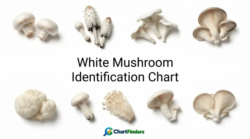
White Mushroom Identification Chart
- Color Is Not Enough: Many of the world’s most lethal mushrooms—including the destroying angel (Amanita virosa and Amanita biscigera)—are pristine white.
- Spore Prints Are Mandatory: Determining the exact color of dropped spores is a non-negotiable step in separating toxic amanitas (white spores) from edible agaricus (brown spores).
- Anatomy Matters: Features hidden beneath the soil line, such as the volva (sac) of an Amanita, must be excavated completely rather than plucked.
- Bruising Reactions: Chemical changes upon cutting, bruising, or handling provide vital diagnostic clues for genus-level separation.
- Habitat Context: Knowing whether a mushroom grows from wood, mycorrhizally with specific trees, or saprobically in lawns drastically narrows down potential species.
To safely navigate the world of white mushrooms, one must rely on structured diagnostic charts rather than superficial resemblances. The following comprehensive matrix categorizes common white wild mushrooms, commercial varieties, and hazardous look-alikes based on their definitive morphological and ecological traits.
| Common Name | Scientific Name | Cap & Surface Traits | Gill Color & Attachment | Stem & Ring Features | Spore Print Color | Edibility & Toxicity |
|---|---|---|---|---|---|---|
| Meadow Mushroom (White Form) | Agaricus campestris | White, smooth to slightly silky; up to 9 cm. | Pink transitioning to dark chocolate brown; free. | Stout white stem with a delicate, fragile skirt-like ring. | Dark chocolate brown | Choice Edible |
| Destroying Angel | Amanita biscigera / virosa | Pristine white, sticky when moist, smooth; up to 12 cm. | Pure white; free from the stem. | White, shaggy/scaly stem with a persistent skirt ring and basal cup (volva). | White | Deadly Toxic (Amatoxins) |
| Yellow-Staining Agaricus | Agaricus xanthodermus | White cap; stains bright yellow immediately when scratched, especially at cap margin and base. | Pinkish then dark brown; free. | Bulbous base, hollow, with a thick skirt ring; stains yellow when bruised. | Dark brown | Toxic (Causes severe gastrointestinal distress) |
| Sweetbread Mushroom (Miller) | Clitopilus prunulus | White to greyish-white, dull, undulating margin; velvety texture. | White turning pinkish; deeply decurrent (running down stem). | Short, off-center to central, solid, white. | Pink | Choice Edible (Caution: do not confuse with white clitocybes) |
| Giant White Funnel / Ivory Funnel | Leucopaxillus giganteus / Clitocybe dealbata | Large (up to 30 cm for Leucopaxillus), white, funnel-shaped with age. | White to cream; decurrent. | Thick, robust, white, lacking a ring or volva. | White | Variable (Leucopaxillus edible; C. dealbata contains muscarine) |
| Spring Polypore / Oyster Look-alike | Lentinus tigrinus or white pleurotoid forms | White to cream, fan-shaped, growing on wood. | White, decurrent on lateral stem. | Off-center to lateral stem or sessile. | White | Edible when young (must verify wood-rotting habit) |
Analyzing this matrix highlights a critical mycological principle: superficial similarities in cap color mask profound differences in internal chemistry and reproductive structures. A thorough exploration of these anatomical and chemical markers is essential for field safety.
Anatomical Markers in White Mushroom Morphology
When studying white mushrooms, general impressions must be replaced by systematic dissection and observation. Because many white fungi share identical coloration, field identification hinges on architectural details across the cap, gills, stem, and base.
Cap (Pileus) Textures and Margins
The cap of a white mushroom provides the first layer of identification data. Foragers must examine:
- Surface Texture: Is the cuticle dry, silky, sticky (viscid), scaly, or smooth? Amanitas often retain soft, cottony universal veil patches on their caps, while some edible Agaricus species develop fine brown fibers as they expand.
- Margin Incurvature: In young specimens, observe whether the margin of the cap is rolled inward (incurved) toward the gills or straight.
- Margin Striations: Check if the edge of the cap is grooved (striate), which is common in certain harmless species but also present in some toxic ones if moisture is high.
Gill (Lamellae) Color Progression and Attachment
Gills are the primary spore-bearing surfaces, and their color changes over time offer some of the most reliable taxonomic separations in the fungal kingdom.
- Color Evolution: Edible field mushrooms (Agaricus) start with pale pink gills that rapidly darken to dark chocolate brown as spores mature. Toxic white look-alikes like Amanita maintain pristine white gills from emergence to decay.
- Attachment Type: Gills may be free (not touching the stem), adnate (attached squarely to the stem), or decurrent (running down the length of the stem). Measuring this attachment accurately requires slicing the mushroom vertically down the center.
Stem (Stipe) Structures: Rings, Bulbs, and Volvas
The stem is the structural anchor of the mushroom and holds critical diagnostic evidence that is easily lost if the specimen is pulled rather than dug.
- The Ring (Annulus): Formed when the partial veil ruptures, the ring can be skirt-like, membranous, cobwebty (cortina), or ephemeral. Note its position near the top of the stem or midway down.
- The Volva (Basal Cup): The universal veil completely envelops developing Amanita species. When the mushroom expands, the bottom of this veil remains as a cup, sac, or fragmented collar at the base of the stem. Because this structure often sits entirely underground in loose leaf litter, pulling a mushroom tears the stem above the volva, destroying the most critical warning sign of a deadly toxic species.
Chemical Spot Tests and Spore Print Analysis
Morphology alone cannot always guarantee safety, particularly when separating closely related species within the genus Agaricus. Mycologists and advanced foragers utilize chemical spot tests and spore prints to confirm identities beyond visual doubt.
Conducting a Spore Print
A spore print reveals the true color of a mushroom’s reproductive units, uninfluenced by the color of the gills themselves.
- Detach the mature cap from the stem.
- Place the cap gill-side down on a piece of contrasting paper (half white, half black, or using glass to test against varied backgrounds).
- Cover the cap with a glass or bowl to prevent air currents from disturbing falling spores.
- Leave undisturbed for 4 to 12 hours, then lift the cap to inspect the deposited spore dust color in natural daylight.
Chemical Reagents for Field and Lab ID
Specific chemical reagents applied to the cap or flesh induce rapid color changes that differentiate edible species from toxic ones:
- Potassium Hydroxide (KOH): A 3% to 10% solution applied to the cap surface helps distinguish yellow-staining toxic Agaricus species (which react yellow with KOH or bruised surfaces) from choice edible meadow mushrooms.
- Melzer’s Reagent: Used microscopically to determine amyloid (turning blue-black) or inamyloid spore reactions, critical for separating certain white spored genera.
- Aniline and Phenol: Historical reagents used to test flesh color reactions in complex agaricoid groups.
Major Families and Genera of White Mushrooms
White mushrooms belong to several distinct fungal families. Recognizing the overarching characteristics of these genera prevents misidentifications.
The Genus Agaricus (The Meadow and Button Group)
The genus Agaricus contains both the cultivated white button mushroom and prized wild edibles like Agaricus campestris. Key identifiers include free pink-to-brown gills, a chocolate-brown spore print, and a distinct ring on the stem. Foragers must exercise caution regarding the yellow-staining group (Agaricus xanthodermus), which causes gastrointestinal illness and exhibits a distinct chemical odor resembling phenol, ink, or creosote when bruised.
The Genus Amanita (The Death Caps and Destroying Angels)
Home to the most lethal fungi on Earth, the genus Amanita includes pristine white species such as Amanita virosa, Amanita biscigera, and white variants of Amanita phalloides. These mushrooms feature pure white caps, pure white free gills, white spores, a distinct membranous ring on the upper stem, and a prominent basal volva. Ingestion of these species results in severe, delayed liver and kidney failure.
The Genus Clitocybe and Related White Funnels
Medium to large white mushrooms with decurrent gills often fall into the family Tricholomataceae or genus Clitocybe. While some are edible (such as Clitopilus prunulus with its pink spore print and mealy odor), many small, white, grass-inhabiting Clitocybe species contain high concentrations of muscarine, causing excessive salivation, sweating, and tearing.
Ecological Context and Habitat Indicators
Fungi do not grow randomly; their appearance is tied to specific ecological niches, nutrient sources, and mycorrhizal partners. Factoring in habitat is a vital safety check in mushroom identification.
Mycorrhizal white mushrooms, such as woodland Amanita species, form symbiotic relationships with the roots of specific trees (including oak, beech, pine, and birch). Finding a white mushroom deep in an undisturbed forest floor under mature hardwood canopy immediately raises the probability of encountering a mycorrhizal toxic species.
Conversely, saprobic white mushrooms feed on decaying organic matter and appear in lawns, pastures, gardens, and compost heaps. Cultivated white button mushrooms and wild meadow mushrooms (Agaricus campestris) thrive in open, grassy fields enriched by livestock manure. Wood-rotting white species (such as oyster mushroom look-alikes) emerge directly from decaying logs, stumps, or standing snags, tying their identification directly to the host tree species.
To visualize the ecological distribution of common white mushroom categories across different habitats, examine the breakdown below:
| Habitat Type | Primary Nutrient Source | Common White Genera Present | Edibility & Safety Profile |
|---|---|---|---|
| Open Grasslands & Pastures | Saprobic / Manure-enriched soil | Agaricus, Chlorophyllum | Mix of choice edibles and toxic look-alikes (verify gill color and bruising). |
| Mature Deciduous Forests | Mycorrhizal tree root symbiosis | Amanita, Tricholoma | High risk of lethal toxins (Destroying angels); exercise extreme caution. |
| Conifer Needle Beds | Mycorrhizal / Humus decay | Clitocybe, Inocybe | Many toxic species containing muscarine or unknown gastrointestinal irritants. |
| Dead Wood, Stumps & Logs | Wood-decay saprobes | Pleurotus, Lentinus, Pluteus | Generally safer if wood substrate is correctly identified; verify spore prints. |
Common Mistakes and Critical Safety Pitfalls
Even experienced foragers occasionally fall victim to cognitive biases or flawed identification habits when evaluating white mushrooms. Recognizing these pitfalls is essential for preventing poisoning.
The “All Grocery Store Mushrooms Are Safe” Fallacy
A common beginner error is assuming that any wild mushroom resembling a standard grocery store white button mushroom is safe to consume. Grocery store mushrooms (Agaricus bisporus) are cultivated in sterile, controlled indoor environments. In the wild, immature destroying angels look remarkably similar to grocery store button mushrooms, sharing the same rounded white cap and pale coloration before the cap fully expands.
Failure to Dig for the Volva
Plucking a mushroom by hand rather than digging it out with a knife or trowel leaves the subterranean bulbous base—and the volva—buried in the soil. Without examining the base for a cup or sac, a toxic Amanita can easily be misidentified as a harmless meadow mushroom.
Ignoring Spore Prints in Favor of Visuals
Visual identification alone is never sufficient for white gilled mushrooms. Skipping the spore print means missing the crucial difference between the dark chocolate-brown spores of an edible Agaricus and the pure white spores of a lethal Amanita.
Frequently Asked Questions
Are all white mushrooms poisonous?
No. Many white mushrooms are delicious edibles, including the meadow mushroom (Agaricus campestris), the miller (Clitopilus prunulus), and white forms of the oyster mushroom (Pleurotus ostreatus). However, the proportion of deadly toxic species among all-white gilled mushrooms is exceptionally high, which is why they demand rigorous verification.
How do I tell an edible meadow mushroom from a destroying angel?
Look at the gills and the base. Meadow mushrooms have pink gills that turn dark chocolate-brown as they mature, and they lack a basal volva cup. Destroying angels have pure white gills that never change color, possess a membranous ring, and always feature a prominent cup-like volva at the base of the stem buried in the soil.
Can I touch a toxic white mushroom like a destroying angel?
Yes. Fungal toxins such as amatoxins and phallotoxins are not absorbed through the intact human skin. Handling a poisonous mushroom is safe as long as you thoroughly wash your hands afterward and ensure no residue is transferred to your mouth or food.
Why is a spore print required for white mushroom identification?
A spore print reveals the definitive color of the mushroom’s spores, which is a fixed taxonomic trait used to separate major families. For example, two mushrooms with identical white caps and white stems can belong to entirely different families—one producing white spores (potentially deadly) and the other producing brown spores (potentially edible).
What should I do if I suspect mushroom poisoning?
Seek emergency medical attention immediately. Do not wait for symptoms to subside. Save any remaining mushroom specimens, including scraps, cooking remnants, or vomitus, in a paper bag or clean container to help toxicologists identify the specific toxin ingested and administer the correct treatment.
Conclusion
Navigating the identification of white mushrooms requires patience, meticulous attention to anatomical detail, and an unwavering commitment to safety protocols. Because some of the world’s most lethal fungi wear an innocent white facade, shortcuts have no place in wild foraging. By mastering the diagnostic markers outlined in this guide—from gill color transitions and spore prints to subterranean volvas and chemical spot tests—foragers can transform uncertainty into confident, scientifically grounded field expertise.