
Parasitic infections remain a significant global health challenge, affecting millions of individuals across both tropical regions and developed nations. The complexity of diagnosing these organisms stems from their diverse lifecycles, morphological similarities, and the variable clinical presentations they elicit in human hosts. For clinical laboratorians, parasitologists, and healthcare providers, a structured, evidence-based approach to parasite identification is essential to prevent misdiagnosis, inappropriate treatment, and chronic morbidity.
Historically, parasite identification relied exclusively on wet-mount microscopy, permanent stained smears, and concentration techniques. While these classical modalities remain foundational, modern clinical diagnostics have evolved. Recent advancements integrate high-throughput molecular diagnostics and next-generation metataxonomics for high-resolution detection of intestinal protists and helminths. Understanding how to navigate an identification chart involves synthesizing visual morphology—such as cyst versus trophozoite dimensions, nuclear characteristics, and ova or larval features—with molecular confirmation.
Explore our detailed breakdown on Wood Grain Identification Chart: Field Comparison Matrix.This master guide explores the structural taxonomy of human parasites, dissects diagnostic modalities, provides detailed morphological breakdowns across major parasite classes, and outlines step-by-step identification methodologies. By mastering these criteria, clinicians and laboratory specialists can achieve precise, reproducible diagnostic accuracy.
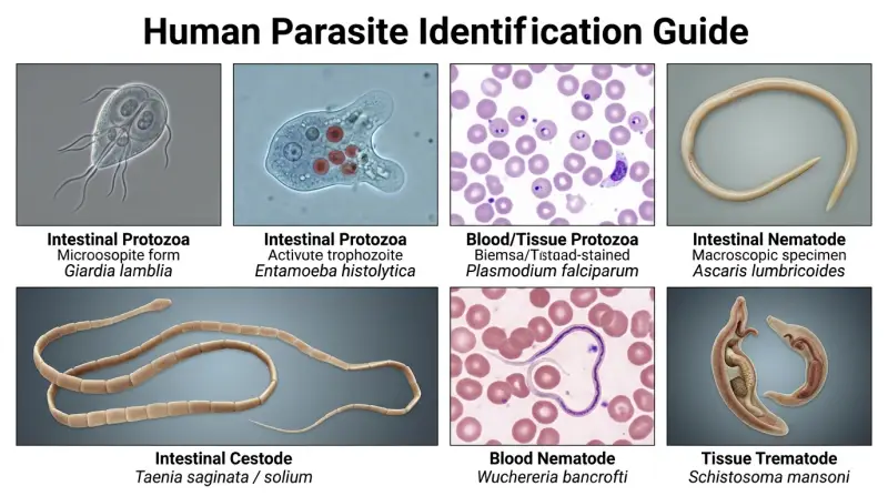
Human Parasite Identification Chart
- Multi-Modal Approach: Modern parasite identification merges classic brightfield/immunofluorescence microscopy with cutting-edge metataxonomics and molecular assays.
- Major Classifications: Human parasites divide primarily into Protozoa (single-celled protists divided into intestinal and blood/tissue types) and Helminths (multicellular worms including nematodes, cestodes, and trematodes).
- Morphological Markers: Diagnostic accuracy depends on recognizing distinct structural markers, such as nuclear karyosomes in amoebic cysts, hooklets in cestode scoleces, and opercula in trematode eggs.
- Staining Precision: Permanent stained smears using trichrome or iron-hematoxylin are non-negotiable for differentiating internal structures of intestinal protozoa.
- Clinical Correlation: Geographic exposure, travel history, and immune status must always accompany laboratory findings to rule out non-pathogenic commensals.
To systematically identify human parasites, laboratories utilize reference charts that group organisms by taxonomic classification, primary specimen source, and diagnostic morphologic features. Below is a comprehensive master reference table outlining the primary medically important human parasites, their morphological hallmarks, and standard diagnostic methodologies.
| Parasite Category | Species Example | Primary Specimen | Diagnostic Stage & Size | Key Morphological Identifiers | Diagnostic Method |
|---|---|---|---|---|---|
| Intestinal Protozoa | Giardia lamblia | Stool | Cyst: 8–14 µm Trophozoite: 9–21 µm | Cyst: 2–4 nuclei, axostyle. Trophozoite: “Falling leaf” motility, two nuclei, sucking disk (“face-like”). | Stool EIA, DFA, trichrome stain, wet mount |
| Intestinal Protozoa | Entamoeba histolytica | Stool | Cyst: 10–20 µm Trophozoite: 12–60 µm | Cyst: 1–4 nuclei with central karyosome; chromatoid bars with blunt ends. Trophozoite: ingested RBCs (pathognomonic), fine peripheral chromatin. | Permanent stained smear, multiplex PCR, stool antigen |
| Blood/Tissue Protozoa | Plasmodium falciparum | Peripheral Blood | Ring form: 1–2 µm Gametocyte: Crescentic | Rings: Multiple per RBC, delicate cytoplasm, double chromatin dots (“accolé”). Schizonts rarely seen in peripheral blood. | Giemsa-stained thin/thick smears, rapid diagnostic tests (RDTs) |
| Intestinal Nematode | Ascaris lumbricoides | Stool | Fertilized Egg: 45–75 µm | Round/oval egg with a thick, golden-brown mammillated (bumpy) albuminous coat surrounding an unsegmented ovum. | Direct saline mount, formalin-ethyl acetate concentration |
| Intestinal Cestode | Taenia saginata / solium | Stool / Proglottids | Egg: 31–43 µm Proglottids vary | Egg: Spherical, thick radially striated brown shell enclosing a hexacanth embryo. Proglottids: Uterine branch counts differentiate species. | Stool concentration, acid-fast stain (for species differentiation), molecular assays |
| Blood Nematode | Wuchereria bancrofti | Nocturnal Blood | Microfilariae: 240–300 µm | Sheathed microfilariae, graceful curves, nuclei do not extend to the very tip of the tail. | Nocturnal thick blood smear, Knott’s technique, PCR |
| Tissue Trematode | Schistosoma mansoni | Stool / Tissue | Egg: 114–180 µm | Large, elongated oval egg featuring a prominent lateral spine near the posterior end; contains a ciliated miracidium. | Kato-Katz thick smear, sedimentation concentration, rectal biopsy |
The table above illustrates the breadth of structural variations encountered across parasite phyla. Interpreting these specifications requires a granular understanding of individual taxonomic groups, their life histories, and the nuances of laboratory specimen processing.
Taxonomic Classification and Biological Architecture
Human parasites are categorized into two major sub-kingdoms: Protozoa (single-celled eukaryotic microorganisms) and Helminths (multicellular invertebrates). Within these broad divisions, specialized morphological features dictate how laboratorians classify and identify each organism.
Intestinal and Urogenital Protozoa
Protozoan parasites alternate between a metabolically active, fragile feeding stage known as the trophozoite and a hardy, environmentally resilient cyst stage. Intestinal protozoa include amoebae, flagellates, ciliates, and coccidia.
- Amoebae (e.g., Entamoeba histolytica, Endolimax nana): Identified primarily by nuclear morphology (karyosome size and chromatin distribution) in permanent stained smears and the presence or absence of ingested erythrocytes.
- Flagellates (e.g., Giardia lamblia, Dientamoeba fragilis): Characterized by locomotory flagella, axostyles, and distinctive pear-shaped (trophozoite) or oval (cyst) outlines.
- Ciliates (e.g., Balantidium coli): The largest human protozoan parasite, recognized by body cilia and a prominent kidney-shaped macronucleus.
- Coccidia (e.g., Cryptosporidium, Cyclospora, Cystoisospora): Intracellular parasites that produce oocysts shed in feces. They require modified acid-fast stains or autofluorescence under UV microscopy due to their small size (4–30 µm).
Blood and Tissue Protozoa
These organisms inhabit the circulatory system and reticuloendothelial tissues. Hemoflagellates (such as Trypanosoma and Leishmania species) are identified by kinetoplasts, undulating membranes, and flagella visible on Giemsa-stained blood or aspirate smears. Apicomplexan blood parasites (such as Babesia and Plasmodium species) parasitize red blood cells, presenting distinct ring forms, trophozoites, schizonts, and gametocytes that require meticulous examination of thin and thick blood films.
Helminths: Nematodes, Cestodes, and Trematodes
Helminthic infections are diagnosed primarily by identifying characteristic ova (eggs), larvae, or adult worm fragments (such as proglottids and scoleces) in clinical specimens.
- Nematodes (Roundworms): Cylindrical, unsegmented worms. Diagnostic forms include distinct eggs (e.g., barrel-shaped eggs with bipolar plugs in Trichuris trichiura) or circulating microfilariae in blood/lymphatics.
- Cestodes (Tapeworms): Ribbon-like, segmented flatworms. Identification relies on examining egg shell striations and hooklets, as well as the morphology of gravid proglottids and the holdfast organ (scolex) equipped with hooks or suckers.
- Trematodes (Flukes): Leaf-shaped, unsegmented flatworms. Their eggs are typically operculated (except for Schistosoma species, which feature distinct terminal or lateral spines) and are recovered in stool, urine, or sputum depending on the species tropism.
Comparative Analysis of Major Parasite Groups
To differentiate clinical presentations, diagnostic approaches, and identification complexities across parasite classes, laboratorians evaluate specific operational parameters. The table below compares intestinal protozoa, blood/tissue protozoa, nematodes, cestodes, and trematodes.
| Parasite Group | Biological Nature | Primary Diagnostic Specimen | Key Structural Hallmarks | Primary Diagnostic Challenges |
|---|---|---|---|---|
| Intestinal Protozoa | Unicellular eukaryotes (Cyst & Trophozoite forms) | Fresh or preserved stool | Nuclear karyosomes, chromatoid bars, flagella, axostyles | Morphologic mimicry between pathogenic and non-pathogenic species |
| Blood & Tissue Protozoa | Intracellular or intravascular single-celled organisms | Peripheral blood, tissue aspirates, biopsy | Ring forms, crescentic gametocytes, kinetoplasts | Intermittent parasitemia, rapid degradation if blood is unprocessed |
| Nematodes (Roundworms) | Multicellular, unsegmented cylindrical worms | Stool, perianal skin (tape test), blood | Thick-shelled eggs, bipolar plugs, sheathed microfilariae | Low egg burdens in light infections; extraintestinal migration stages |
| Cestodes (Tapeworms) | Segmented ribbon-like flatworms | Stool, proglottids | Hexacanth embryos, radially striated shells, scolex hooklets/suckers | Distinguishing species via proglottid uterine branches or molecular assays |
| Trematodes (Flukes) | Leaf-shaped unsegmented flatworms | Stool, urine, sputum, tissue | Operculate eggs, terminal or lateral spines, ciliated miracidia | Specimen specificity (e.g., urine for S. haematobium, sputum for Paragonimus) |
Diagnostic Methodologies: From Microscopy to Metataxonomics
Accurate parasite identification is contingent upon proper specimen collection, timing, preservation, and the application of appropriate analytical techniques. Over-reliance on a single diagnostic method frequently leads to false negatives due to intermittent shedding or low parasite burdens.
Microscopic Examination Techniques
Microscopy remains the of diagnostic parasitology. Laboratories employ several preparation steps:
- Direct Wet Mounts: Useful for detecting motile trophozoites in fresh, warm diarrheal stool specimens. Saline mounts reveal motility, while iodine mounts highlight nuclear structures and glycogen vacuoles in cysts.
- Concentration Procedures: Formalin-ethyl acetate sedimentation and zinc sulfate flotation concentrate ova, cysts, and larvae, significantly increasing detection sensitivity in formed or semi-formed stools.
- Permanent Stained Smears: Trichrome and iron-hematoxylin stains are mandatory for confirming intestinal protozoa. They allow detailed visualization of nuclear chromatin, karyosomes, and cytoplasmic inclusions that cannot be discerned on wet mounts.
- Specialized Stains: Modified acid-fast staining is essential for identifying oocysts of coccidian parasites (Cryptosporidium, Cyclospora, Cystoisospora), which stain variably red against a blue background. Calcofluor white fluorescence is utilized for microsporidia and fungal elements.
Molecular and Advanced Metataxonomic Diagnostics
As diagnostic protocols evolve, molecular testing has transformed clinical parasitology. Multiplex real-time polymerase chain reaction (PCR) assays offer superior sensitivity and specificity compared to traditional microscopy, distinguishing pathogenic species (such as Entamoeba histolytica) from morphologically identical non-pathogenic commensals (such as Entamoeba dispar).
, the integration of metataxonomics—high-throughput amplicon sequencing of marker genes (such as the 18S rRNA gene for protists and ITS regions for helminths)—enables comprehensive microbiome and parasitome profiling. Metataxonomics excels in complex coinfection screening, epidemiological surveillance, and diagnosing low-level shedding where microscopic visualization fails.
Step-by-Step Parasite Identification Workflow
When presented with an unknown clinical specimen, laboratory personnel follow a rigorous, sequential decision-making framework to ensure accurate identification.
| Workflow Step | Operational Action | Primary Objective | Key Pitfall to Avoid |
|---|---|---|---|
| 1. Clinical & Travel Intake | Review patient travel history, exposure risks, and clinical symptoms. | Narrow differential diagnosis (e.g., suspecting malaria in returning travelers). | Ignoring travel history, leading to missed exotic pathogens. |
| 2. Specimen Processing | Select appropriate fixative (e.g., 10% formalin, PVA) and perform concentration. | Optimize recovery of low-abundance ova, cysts, or larvae. | Over-centrifugation or delayed fixation, destroying fragile trophozoites. |
| 3. Screening Microscopy | Examine wet mounts and thick/thin blood films under low (10x) and high-dry (40x) power. | Rapidly locate large helminth eggs, microfilariae, or malarial parasites. | Rushing the slide review and missing low-burden infections. |
| 4. Confirmatory Staining | Review trichrome, iron-hematoxylin, or acid-fast stained slides under oil immersion (100x). | Evaluate internal nuclear structure, inclusion bodies, and oocyst staining properties. | Misidentifying fecal artifacts (macrophages, yeast, pollen) as protozoan cysts. |
| 5. Molecular Validation | Perform multiplex PCR or metataxonomic sequencing on ambiguous isolates. | Definitive species-level differentiation and resolution of morphologic mimics. | Relying solely on molecular panels without correlating viable organism shedding. |
Common Diagnostic Mistakes and Artifact Pitfalls
Even experienced microscopists encounter diagnostic pitfalls. Recognizing artifacts and morphological look-alikes is critical to avoiding misdiagnosis and unnecessary therapeutic interventions.
Fecal Artifacts Versus True Parasites
Stool specimens contain numerous non-parasitic structures that mimic ova and cysts. Pollen grains can resemble nematode or trematode eggs due to their thick walls and internal patterning, but they lack distinct cellular embryos or opercula. Vegetable cells, starch granules, and plant hairs are frequently mistaken for helminth larvae or worm fragments. Similarly, white blood cells (macrophages and neutrophils) in dysenteric stools closely resemble amoebic trophozoites; permanent trichrome staining is required to reveal nuclear details and distinguish leukocytes from pathogens.
Species Mimicry
Morphological identification is severely limited when organisms share identical microscopic appearances. The classic example is the Entamoeba complex: Entamoeba histolytica (pathogenic) is morphologically indistinguishable from Entamoeba dispar and Entamoeba moshkovskii (non-pathogenic) under standard light microscopy. Conclusive identification mandates antigen detection or molecular assays (PCR), reinforcing the necessity of multi-modal diagnostic pathways.
Frequently Asked Questions
A combination of permanent stained smears (using trichrome or iron-hematoxylin) and molecular assays (such as multiplex real-time PCR) provides the highest sensitivity and specificity. While concentration wet mounts catch many cysts, permanent staining is essential for visualizing internal nuclear characteristics required for definitive protozoan identification.
Many intestinal parasites (such as Giardia lamblia and various helminths) shed cysts, ova, or larvae intermittently rather than continuously. Collecting three stool specimens over a 7- to 10-day period significantly increases diagnostic yield and reduces false-negative results caused by low or fluctuating parasite burdens.
While RDTs (such as those detecting malaria antigens like HRP-2 or pLDH) are invaluable for rapid point-of-care screening, they do not replace microscopic examination or molecular testing. Microscopy (thick and thin Giemsa-stained smears) remains mandatory to confirm species, quantify parasitemia levels, and monitor therapeutic response.
Metataxonomics utilizes high-throughput sequencing of specific marker regions (e.g., 18S rRNA) to profile complex communities of protists and helminths simultaneously. This approach overcomes the limitations of observer subjectivity, resolves morphologically identical species complexes, and detects low-abundance coinfections that standard microscopy frequently overlooks.
True parasite eggs possess consistent, biologically structured features such as specialized shells (e.g., opercula in trematodes, mammillated coats in Ascaris, or distinct radial striations in Taenia) and organized internal cellular content (embryos or blastomeres). Artifacts such as pollen grains, vegetable spirals, and fat globules lack these ordered internal structures and exhibit variable staining characteristics.
Conclusion
Mastering human parasite identification requires a disciplined integration of classical morphology, advanced staining techniques, molecular testing, and clinical context. As diagnostic technologies evolve to incorporate high-throughput metataxonomics and multiplex PCR, laboratory professionals must maintain a deep understanding of fundamental structural markers—from protozoan nuclear karyosomes to helminthic egg spines. By adhering to structured identification workflows and remaining vigilant against common artifact pitfalls, healthcare laboratories can ensure rapid, accurate diagnoses that directly improve patient outcomes.